AVMs and cavernomas
Abnormal tangles or clusters of blood vessels in the brain. Both can bleed, and both are often found before they do, which is when the decisions are hardest and most worth making carefully.
Greenslopes Private Hospital, Brisbane
Watkins Medical Centre, Spring Hill, or telehealth
A current GP or specialist referral is needed
At a glance
Arteries connected directly to veins without the usual small vessels between.
High pressure; can bleed, cause seizures or headache.
A cluster of thin-walled, low-pressure vessels, like a small berry.
Small bleeds; seizures; often watched.
Observation, microsurgery, embolisation, radiosurgery.
Often a combination for AVMs.
Progressive narrowing of the brain’s main arteries.
Treated with bypass surgery using scalp vessels.
What these malformations are
Both are present from early life rather than acquired. They differ in pressure, in how they bleed, and in how they are treated.
An arteriovenous malformation (AVM) is a tangle in which arteries connect straight into veins. Blood passes through under arterial pressure, the vessel walls are abnormal, and the malformation can bleed into the brain. AVMs are graded on their size, their position relative to important brain areas, and how they drain, and the grade is a guide to how safely they can be removed.
A cavernoma (cavernous malformation) is a cluster of thin-walled vessels with sluggish, low-pressure flow. It bleeds in small amounts rather than catastrophically, but repeated small bleeds can cause seizures or, in the brainstem, progressive symptoms. Some people have several cavernomas, and in some families they are inherited. A developmental venous anomaly is often found beside a cavernoma; it is a normal variant and is left alone.
Moyamoya disease is different again: a gradual narrowing of the large arteries at the base of the brain, which the brain tries to bypass with a fine network of fragile vessels. It causes strokes or bleeds, in children and adults, and is treated by surgery that brings a new blood supply from the scalp.
Symptoms
Many AVMs and cavernomas cause no symptoms until they bleed, or are found on a scan done for another reason. When they do cause symptoms, these are the common ones.
- A bleed: sudden headache, weakness, numbness, speech or vision disturbance, or loss of consciousness
- Seizures, sometimes the first sign of either condition
- Headache that is persistent or unusual for you
- Gradually progressive weakness, numbness or double vision, particularly with a brainstem cavernoma
- In moyamoya: brief episodes of weakness or speech difficulty, or a stroke, in a young person
A sudden severe headache, new weakness, confusion, drowsiness or a seizure needs an emergency department, not a website. Call 000.
How it is diagnosed
MRI shows cavernomas clearly and is the main test for them; they do not show on angiography. For an AVM, MRI is followed by CT or MR angiography and then a catheter angiogram, which maps the feeding arteries and draining veins in the detail needed to plan treatment. Moyamoya is diagnosed on angiography and perfusion imaging.
The scan is on the screen, the options are set out on equal terms, and nothing is booked until you have had time to think.
Treatment options
For an AVM the aim of treatment is to remove it completely, because a partly treated AVM can still bleed. For a cavernoma the question is whether it is causing enough trouble to justify an operation.
- ObservationInterval MRI for cavernomas that have not bled or caused seizures, and for some AVMs where the risks of treatment are high. Seizures, where present, are managed with a neurologist.
- Microsurgical removalCraniotomy to remove the malformation under the operating microscope with neuronavigation. For an accessible AVM or a symptomatic cavernoma, complete removal ends the risk of bleeding from it.
- EmbolisationGlue or coils delivered through a catheter by an interventional neuroradiologist to block feeding vessels. Usually a step before surgery or radiosurgery for an AVM; occasionally a treatment on its own.
- RadiosurgeryFocused radiation for small or deep AVMs that are hard to reach, planned with a radiation oncologist. The vessels close over two to three years, during which the risk of bleeding remains.
- Bypass surgeryFor moyamoya disease, an operation that connects a scalp artery to the surface of the brain so that a new blood supply grows in over the following months.
Not every option applies to every person. Which ones apply to you, and in what order, is explained at the consultation and confirmed in writing before anything is booked.
Recovery and follow-up
Recovery depends on whether there has been a bleed, on the position of the malformation and on which treatment was used.
After a craniotomy, several days in hospital and some weeks of tiredness are usual. After removal of an AVM, an angiogram confirms that nothing remains. Seizure medication, where it was started, is continued for a period and then reviewed with the neurologist; driving rules apply after any seizure.
Cavernomas that are being watched are imaged at intervals, sooner if symptoms change. Where a cavernoma has been removed, a follow-up MRI confirms the result. After bypass surgery for moyamoya, imaging over the following months shows the new blood supply establishing itself.
Before you decide
The decision to treat an AVM or cavernoma that has never bled is one of the more finely balanced in neurosurgery. The consultation goes through the estimated risk of bleeding over your lifetime, the risks of each treatment, and what watching would involve, and there is time to think.
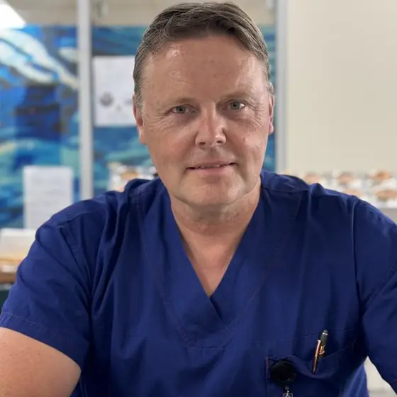
Dr Gert TollessonNeurosurgeon, Brisbane
Ready to talk it through?
A current referral is needed to book. If you do not have one yet, call the rooms on (07) 3870 3708 and they will explain what to ask your GP for.
Request a consultation
Send your details and the rooms will call you to arrange a time. Referrals are triaged as they arrive.
Monday to Friday, 8:30am to 4:30pm. No referral yet? The rooms will explain what to ask your GP for.
Before your appointment
What happens from referral to follow-up, and what it costs.