Meningioma and skull base tumours
Tumours that arise from the membranes covering the brain, or from the bone and tissue at the base of the skull. Most are benign and slow-growing, and many need nothing more than watching.
Greenslopes Private Hospital, Brisbane
Watkins Medical Centre, Spring Hill, or telehealth
A current GP or specialist referral is needed
At a glance
Meningioma, acoustic neuroma (vestibular schwannoma), chordoma and other skull base tumours.
Usually benign; meningiomas are graded 1 to 3.
MRI with contrast; CT for bone detail; hearing tests for acoustic neuroma.
Often found by chance on a scan done for another reason.
Observation, surgery, or radiosurgery.
Chosen on size, position, growth and symptoms.
Greenslopes Private Hospital (adults).
Queensland Children’s Hospital (children).
What these tumours are
Meningiomas are the most common primary tumour inside the skull. Skull base tumours are rarer, and their position next to nerves and blood vessels is what makes them a specialist problem.
A meningioma grows from the arachnoid, one of the membranes that wrap the brain and spinal cord. It sits on the surface of the brain and pushes on it rather than growing into it. Most are grade 1, meaning benign and slow. A smaller number are grade 2 (atypical) or grade 3 (malignant) and behave more aggressively. Meningiomas are more common in women and become more common with age.
Skull base tumours arise from the bone, nerves and tissues at the floor of the skull. The most common is the acoustic neuroma, a benign tumour on the hearing and balance nerve. Others include chordomas, pituitary tumours (which have their own page) and meningiomas of the skull base itself.
What all of these have in common is neighbours. The base of the skull carries the nerves for vision, eye movement, facial sensation and movement, hearing, swallowing and speech, and the main arteries to the brain. A tumour’s position relative to those structures shapes every decision about it.
Symptoms
Many of these tumours cause no symptoms and are found on a scan done for something else. When symptoms occur they reflect the tumour’s position.
- Headache, or seizures, from a tumour pressing on the brain surface
- Gradual hearing loss, ringing or imbalance on one side (acoustic neuroma)
- Blurred or double vision, or loss of part of the visual field
- Facial numbness, pain or weakness
- Weakness or clumsiness of a limb, or changes in speech
- Changes in personality or concentration from a tumour at the front of the brain
An incidental finding is common. Being told a scan has shown a meningioma is alarming, but it is often the start of a plan to watch rather than to operate.
How it is diagnosed
MRI with contrast shows the tumour and its relationship to the brain, nerves and vessels; CT shows the bone of the skull base. Hearing and balance tests are part of the assessment for an acoustic neuroma, and visual field testing where the optic pathways are close. Tissue diagnosis comes from the operation, where one is done; a plan to watch does not need it.
The scan is on the screen, the options are set out on equal terms, and nothing is booked until you have had time to think.
Treatment options
Three questions decide the plan: is the tumour causing symptoms, is it growing, and what would it cost to remove it safely. ENT, radiation oncology and neuroradiology colleagues are involved where the position calls for it.
- ObservationInterval MRI, usually at 6 to 12 months and then yearly, for small tumours that are not causing symptoms. Many meningiomas never need anything else.
- SurgeryCraniotomy or a skull base approach to remove the tumour under the operating microscope with neuronavigation. The aim is complete removal where it is safe; where a nerve or vessel is at risk, a deliberate partial removal with later radiosurgery can be the better plan.
- RadiosurgeryFocused radiation (Gamma Knife or LINAC) for small tumours, for tumour left behind at surgery, or for regrowth, planned with a radiation oncologist. No incision, but the effect is gradual.
- Endoscopic surgerySome tumours at the front of the skull base can be reached through the nose with an endoscope, with an ENT surgeon, avoiding a craniotomy.
- RadiotherapyA fractionated course for grade 2 or 3 meningiomas, or where the tumour is too large for radiosurgery.
Not every option applies to every person. Which ones apply to you, and in what order, is explained at the consultation and confirmed in writing before anything is booked.
Recovery and follow-up
After surgery, recovery takes weeks; follow-up takes years, because these tumours can regrow slowly and quietly.
A hospital stay of several days is usual after a craniotomy, longer for complex skull base operations. Tiredness, headache and scalp discomfort settle over weeks. The pathology result confirms the grade and guides whether any further treatment is needed.
Follow-up MRI is arranged at intervals, typically at three to six months after surgery and then yearly for a number of years. For an acoustic neuroma, hearing and facial nerve function are reviewed alongside the scans.
Watching a meningioma
A plan to watch means an MRI at an agreed interval, a clear description of what would change the advice, and a way to contact the rooms sooner if symptoms appear. It is an active decision, and it is reviewed at each scan.
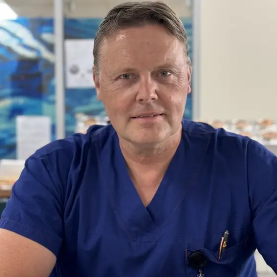
Dr Gert TollessonNeurosurgeon, Brisbane
Ready to talk it through?
A current referral is needed to book. If you do not have one yet, call the rooms on (07) 3870 3708 and they will explain what to ask your GP for.
Request a consultation
Send your details and the rooms will call you to arrange a time. Referrals are triaged as they arrive.
Monday to Friday, 8:30am to 4:30pm. No referral yet? The rooms will explain what to ask your GP for.
Before your appointment
What happens from referral to follow-up, and what it costs.